Oral Submucous Fibrosis
Oral Submucous Fibrosis
At Dr. Dahiwals Facial Aesthetics Center & Dental Clinic, we specialize in providing comprehensive care for oral submucous fibrosis. Our services include:
Oral submucous fibrosis (OSF) is a chronic, complex, premalignant (1% transformation risk) condition of the oral cavity, characterized by juxta-epithelial inflammatory reaction and progressive fibrosis of the submucosal tissues (the lamina propria and deeper connective tissues). As the disease progresses, the oral mucosa becomes fibrotic to the point that the person is unable to open the mouth. The condition is remotely linked to oral cancers and is associated with the chewing of areca nut and/or its byproducts, commonly practiced in South and South-East Asian countries. The incidence of OSF has also increased in western countries due to changing habits and population migration.
Epidemiology
The incidence of the disease is higher in people from certain parts of the world including South and South East Asian, South Africa and the Middle Eastern countries.
Symptoms
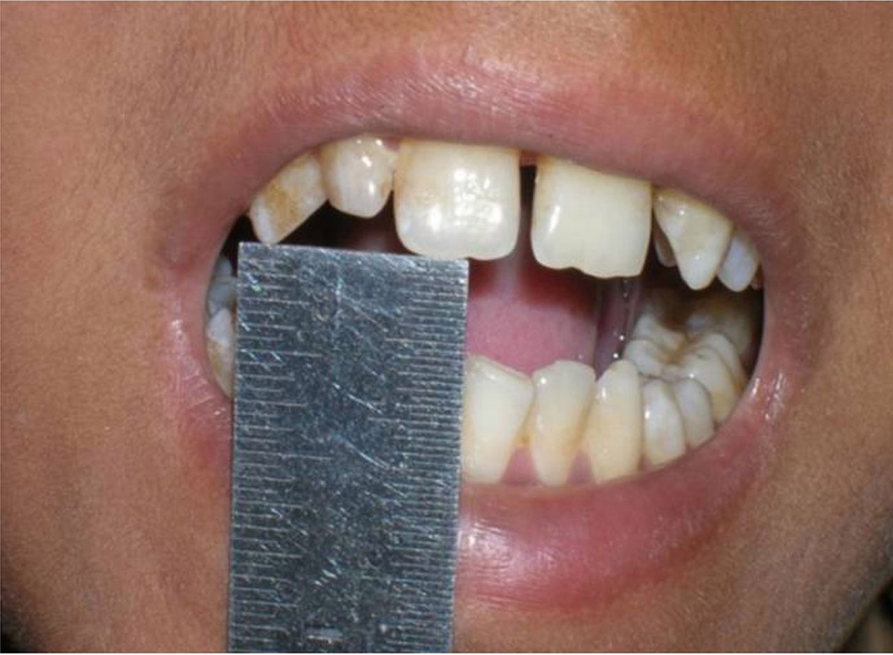
In the initial phase of the disease, the mucosa feels leathery with palpable fibrotic bands. The oral mucosa loses resiliency in the advanced stage and becomes blanched and stiff. This blanched and stiff mucosa is considered to lead to a progressive reduction in mouth opening but seems to be an oversimplification of the pathology. The degree of mouth opening is also determined by the severity of oral symptoms, such as recurring or persistent glossitis and stomatitis, a fact that many researchers ignore. This phenomenon is explained by the term Reflectory Trismus, where the above symptoms dictate the degree of mouth opening through activation of the 5th and 9th cranial nerves. However, muscle damage and fibrosis play a larger contributory role.[9] The condition is believed to begin in the posterior part of the oral cavity and gradually spread outward. The premise posterior to the anterior progression of oral submucous fibrosis has been recently rebutted based on several reports stating that the disease may be restricted to the anterior part of the oral cavity without involvement of posterior parts; the sites are dictated by the manner of use anterior areas of the oral cavity when spitting and posterior when swallowed.
Other features of the disease include:
• Xerostomia
• Recurrent ulceration
• Pain in the ear or deafness
• Nasal intonation of voice
• Restriction of the movement of the soft palate
• A budlike shrunken uvula
• Thinning and stiffening of the lips
• Pigmentation of the oral mucosa
• Dryness of the mouth and burning sensation (stomatopyrosis)
• Decreased tongue protrusion
Cause
• Dried products such as paan masala and gutkha have higher concentrations of areca nut and appear to cause the disease.
• Areca nut is the definitive causative agent of OSF.
• A new term was introduced by Sharma et al., in 2024 “Areca Nut induced oral Fibrosis (AIOF)” since fibrosis in the oral cavity can occur due to a variety of causes.
• Fibrosis due to other causes has distinctive features and should be named according to cause and not called oral submucous fibrosis
o Immunological diseases can occur in the background of graft vs Host Disease or as an oral manifestation of systemic sclerosis
o Khat Induced Oral fibrosis
o Oral fibrosis due to Iron defiency anemia
o Tobacco-induced oral fibrosis
Treatment
Biopsy screening, although necessary, is not mandatory; most dentists can visually examine the area and proceed with the proper course of treatment.
Treatment includes:
• Abstention from chewing areca nut (also known as betel nut) and tobacco
• Minimizing consumption of spicy foods, including chiles
• Maintaining proper oral hygiene
• Supplementing the diet with foods rich in vitamins A, B complex, and C and iron
• Forgoing hot fluids like tea, coffee
• Forgoing alcohol
• Employing a dental surgeon to round off sharp teeth and extract third molars
• Interprofessional treatment approach
Treatment also includes following:
• The prescription of chewable pellets of hydrocortisone (Efcorlin); one pellet to be chewed every three to four hours for three to four weeks
• 0.5 ml intralesional injection Hyaluronidase 1500 IU mixed in 1 ml of Lignocaine into each buccal mucosa once a week for 4 weeks or more as per condition
• 0.5 ml intralesional injection of Hyaluronidase 1500 IU and 0.5 ml of injection Hydrocortisone acetate 25 mg/ml in each buccal mucosa once a week alternatively for 4 weeks or more as per condition
• Submucosal injections of hydrocortisone 100 mg once or twice daily depending upon the severity of the disease for two to three weeks
• Submucosal injections of human chorionic gonadotrophins (Placentrax) 2–3 ml per sitting twice or thrice in a week for three to four weeks
• Surgical treatment is recommended in cases of progressive fibrosis when interincisor distance becomes less than 2 centimetres (0.79 in). (Multiple release incisions deep to mucosa, submucosa and fibrotic tissue and suturing the gap or dehiscence so created by mucosal graft obtained from tongue and Z-plasty. In this procedure multiple deep z-shaped incisions are made into fibrotic tissue and then sutured in a straighter fashion.)
• Pentoxifylline (Trental), a methylxanthine derivative that has vasodilating properties and increases mucosal vascularity, is also recommended as an adjunct therapy in the routine management of oral submucous fibrosis.
• IFN-gamma is an antifibrotic cytokine which alters collagen synthesis and helps in OSF.
• Colchicine tablets 0.5 mg twice a day
• Lycopene, 16 mg a day helps in improvement of OSF
The treatment of patients with oral submucous fibrosis depends on the degree of clinical involvement. If the disease is detected at a very early stage, cessation of the habit is sufficient. Most patients with oral submucous fibrosis present with moderate-to-severe disease. Severe oral submucous fibrosis is irreversible. Moderate oral submucous fibrosis is reversible with cessation of habit and mouth opening exercise. Current modern day medical treatments can make the mouth opening to normal minimum levels of 30 mm mouth opening with proper treatment
FAQ
OSMF is a chronic, potentially debilitating condition characterized by the progressive fibrosis of the oral mucosa, leading to restricted mouth opening and other oral complications. It is commonly associated with betel nut or tobacco chewing.
Symptoms may include difficulty in opening the mouth, a burning sensation in the mouth, reduced tongue movement, altered taste sensation, and the presence of fibrous bands or ulcers in the oral cavity.
Diagnosis typically involves a thorough clinical examination by a qualified dentist or oral healthcare professional. In some cases, additional tests such as biopsy or imaging studies may be required to confirm the diagnosis and assess the extent of the condition.
Yes, with early diagnosis and appropriate intervention, the progression of OSMF can be halted, and symptoms can be managed effectively. Treatment approaches may vary depending on the severity of the condition and may include medication, lifestyle modifications, oral exercises, and surgical interventions in advanced cases.