Oral lichen planus and lichenoid reaction
Oral lichen planus and lichenoid reaction
Welcome to Dr. Dahiwals Facial Aesthetics Center & Dental Clinic, where we specialize in the treatment of oral lichen planus and lichenoid reaction. Our comprehensive services are designed to provide effective solutions for these conditions, ensuring optimal oral health and overall well-being.
Lichen planus (LP) is a chronic inflammatory and autoimmune disease that affects the skin, nails, hair, and mucous membranes. It is not an actual lichen, but is named for its appearance. It is characterized by polygonal, flat-topped, violaceous papules and plaques with overlying, reticulated, fine white scale (Wickham’s striae), commonly affecting dorsal hands, flexural wrists and forearms, trunk, anterior lower legs and oral mucosa. The hue may be gray-brown in people with darker skin. Although there is a broad clinical range of LP manifestations, the skin and oral cavity remain as the major sites of involvement. The cause is unknown, but it is thought to be the result of an autoimmune process with an unknown initial trigger. There is no cure, but many different medications and procedures have been used in efforts to control the symptoms.
The term lichenoid reaction (lichenoid eruption or lichenoid lesion) refers to a lesion of similar or identical histopathologic and clinical appearance to lichen planus (i.e., an area which resembles lichen planus, both to the naked eye and under a microscope). Sometimes dental materials or certain medications can cause lichenoid reactions. They can also occur in association with graft versus host disease
Lichen planus lesions are so called because of their “lichen-like” appearance and can be classified by the site they involve, or by their morphology.
Lichen planus may be categorized as affecting mucosal or cutaneous surfaces.
• Cutaneous forms are those affecting the skin, scalp, and nails.
• Mucosal forms are those affecting the lining of the gastrointestinal tract (mouth, pharynx, esophagus, stomach, anus), larynx, and other mucosal surfaces including the genitals, peritoneum, ears, nose, bladder and conjunctiva of the eyes.
Signs and symptoms
• Lichen planus affecting the lower lip
• Although lichen planus can present with a variety of lesions, the most common presentation is as a well defined area of purple-coloured, itchy, flat-topped papules with interspersed lacy white lines (Wickham’s striae). This description is known as the characteristic “6 Ps” of lichen planus: planar (flat-topped), purple, polygonal, pruritic, papules, and plaques. This rash, after regressing, is likely to leave an area of hyperpigmentation that slowly fades. That said, a variety of other lesions can also occur.
Lichen planus affecting mucosal surfaces may have one lesion or be multifocal. Examples of lichen planus affecting mucosal surfaces include:
• Esophageal lichen planus, affecting the esophageal mucosa. This can present with difficulty or pain when swallowing due to oesophageal inflammation, or as the development of an esophageal stricture. It has also been hypothesized that it is a precursor to squamous cell carcinoma of the esophagus.
• Genital lichen planus, which may cause lesions on the glans penis or skin of the scrotum in males, and the vulva or vagina in females. Symptoms may include lower urinary tract symptoms associated with stenosis of the urethra, painful sexual intercourse, and itching.[10] In females, Vulvovaginal-gingival syndrome, is severe and distinct variant affecting the vulva, vagina, and gums, with complications including scarring, vaginal stricture formation,[36] or vulva destruction. The corresponding syndrome in males, affecting the glans penis and gums, is the peno-gingival syndrome. It is associated with HLA-DQB1.
Mouth
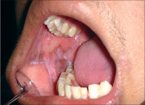
Classic white striations of non-erosive lichen planus in the left buccal mucosa (left cheek)
Oral lichen planus (also termed oral mucosal lichen planus), is a form of mucosal lichen planus, where lichen planus involves the oral mucosa, the lining of the mouth. This may occur in combination with other variants of lichen planus. Six clinical forms of oral lichen planus (OLP) are recognized:
Lesion morphology Description
Reticular The most common presentation of oral lichen planus (OLP), is characterised by the net-like or spider web-like appearance of lacy white lines, oral variants of Wickham’s straiae.This is usually asymptomatic. Reticular OLP may eventually progress to the more severe subtypes such as the erosive form.
Erosive/ Ulcerative The second most common form and the most advance form of oral lichen planus, is characterised by oral ulcers presenting with persistent, irregular areas of redness, ulcerations and erosions covered with a yellow slough. This can occur in one or more areas of the mouth. In 25% of people with erosive oral lichen planus, the gums are involved, described as desquamative gingivitis (a condition not unique to lichen planus). This may be the initial or only sign of the condition. Involvement of the dorsum of the tongue might cause an altered sense of taste (dysgeusia).
Papular This form is characterized by small white pinpoint papules that are asymptomatic. Hence, they can be easily missed during a routine checkup. It is referred to as the initial and transient phase of OLP.
Plaque-like Large, homogenous white patches are characteristic of this form which may resemble leukoplakia. This form is more prevalent in tobacco smokers.
Atrophic This form is a common presentation that has similarities to the erosive form. It has a more prominent atrophic lesion on a background of erythema with radiating white striae at the margins. Atrophic oral lichen planus may also manifest as desquamative gingivitis.
Bullous Rare form of OLP characterized by fluid-filled vesicles ranging in size from 1 to 2 mm to several cm in diameter. The vesicles or bullae appear white or gray-purple in color and are fluctuant. The fluid in the vesicles are usually clear but may be hemorrhagic or purulent upon secondary infection. These rupture easily and they leave an ulcerated, painful surface.[citation needed]
These types often coexist in the same individual. Oral lichen planus (OLP) tends to present bilaterally as mostly white lesions on the inner cheek, although any mucosal site in the mouth may be involved. Other sites, in decreasing order of frequency, may include the tongue, lips, gingivae, floor of the mouth, and very rarely, the palate.
Generally, oral lichen planus tends not to cause any discomfort or pain, although some people may experience soreness when eating or drinking acidic or spicy foodstuffs or beverages. When symptoms arise, they are most commonly associated with the atrophic and ulcerative subtypes. These symptoms can include a burning sensation to severe pain. They may also experience mucosal bleeding in response to mild trauma, such as toothbrushing. Additionally, the Koebner phenomenon (the development of new lesions at sites of trauma) is not only present in cutaneous lichen planus (CLP) but can also occur in the setting of OLP.
A diagnosis of oral lichen planus (LP) is confirmed through review of the patient history, physical examination, and histologic findings.[citation needed]
The clinical evaluation should include a patient history that assesses the following:
• History of LP involving other body sites or other skin disorders that may present with similar findings (e.g., autoimmune blistering diseases)
• Presence of associated symptoms (e.g., pain, burning)
• Medication the patients are taking within the few weeks to months after drug initiation e.g. antihypertensives, antidepressants, diuretics, antidiabetics, NSAIDs, etc. to evaluate for the possibility of an oral lichenoid drug eruption
• History of dental restorations, use of dental appliances, or oral exposure to substances that may cause oral lichenoid contact eruptions (e.g. dental composites, cobalt chromium based dentures etc.)
Treatment
There is no cure for lichen planus, and so treatment of cutaneous and oral lichen planus is for symptomatic relief or due to cosmetic concerns. When medical treatment is pursued, first-line treatment typically involves either topical or systemic corticosteroids, and removal of any triggers. Without treatment, most lesions will spontaneously resolve within 6–9 months for cutaneous lesions, and longer for mucosal lesions.
Reassurance that the condition is benign, elimination of precipitating factors and improving oral hygiene are considered initial management for symptomatic OLP, and these measures are reported to be useful. Treatment usually involves topical corticosteroids (such as betamethasone, clobetasol, dexamethasone, and triamcinolone) and analgesics, or if these are ineffective and the condition is severe, then systemic corticosteroids may be used. Calcineurin inhibitors (such as pimecrolimus, tacrolimus or cyclosporin) are sometimes used. While topical steroids are widely accepted as first line treatment for mucosal lichen planus, there is only weak evidence to support their effectiveness for erosive oral lichen planus.
FAQ
Oral lichen planus and lichenoid reaction are chronic inflammatory conditions that affect the mucous membranes in the mouth, causing discomfort, pain, and other oral symptoms.
Symptoms may include white, lacy patches or lesions in the mouth, soreness or burning sensation, difficulty swallowing, and sensitivity to certain foods or beverages.
The exact cause is unknown, but factors such as autoimmune reactions, certain medications, viral infections, and allergic reactions may contribute to the development of these conditions.
Treatment aims to manage symptoms and control inflammation. This may involve medications such as corticosteroids, immunosuppressants, or topical agents, along with lifestyle modifications and oral hygiene practices to promote healing and relieve discomfort.