Erythroplakia
Erythroplakia Treatment:
At Dr. Dahiwals Facial Aesthetics Center & Dental Clinic, we offer comprehensive and effective treatment for erythroplakia, a potentially serious condition characterized by red patches in the mouth that may indicate precancerous or cancerous lesions. Our experienced team is dedicated to providing personalized care and advanced treatment options to address erythroplakia and promote oral health.
Erythroplakia is a clinical term to describe any erythematous (red) area on a mucous membrane, that cannot be attributed to any other pathology.
The term erythroplasia was coined by Louis Queyrat to describe a precancerous red lesion of the penis. This gave rise to the term erythoplasia of Queyrat. Depending upon the context, this term may refer specifically to carcinoma in situ of the glans penis or vulva appearing as a red patch, or may be used as a synonym of erythroplasia on other mucous membrane or transitional sites.
It mainly affects the glans penis (the head of the penis), although uncommonly it may present on the mucous membranes of the larynx, and rarely, the mouth, or the anus.
Erythroplakia is analogous to the term leukoplakia which describes white patches. Together, these are the 2 traditionally accepted types of premalignant lesion in the mouth, When a lesion contains both red and white areas, the term “speckled leukoplakia” or “eyrthroleukoplakia” is used.
Although oral erythroplakia is much less common than leukoplakia, erythroplakia carries a significantly higher risk of containing dysplasia or carcinoma in situ, and of eventually transforming into invasive squamous cell carcinoma (a type of oral cancer).
Signs and symptoms
- Although often the terms erythroplasia and erythroplakia are used synonymously, some sources distinguish them, stating that the latter is macular (flat) while the former is papular (bumpy).
- Erythroplakia of the genital mucosae is often referred to as erythroplasia of Queyrat.
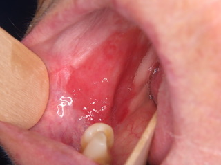
- The most common areas in the mouth where erythroplakia is found are the floor of the mouth, buccal vestibule, the tongue, and the soft palate. It appears as a red macule or plaque with well-demarcated borders. The texture is characterized as soft and velvety. An adjacent area of leukoplakia may be found along with the erythroplakia.
- Erythroplasia may also occur on the laryngeal mucosa, or the anal mucosa.
Causes
Erythroplakia has an unknown cause but researchers presume it to be similar to the causes of squamous cell carcinoma. Carcinoma is found in almost 40% of erythroplakia. It is mostly found in elderly men around the ages of 65 – 74. It is commonly associated with smoking.
Alcohol and tobacco use have been described as risk factors.
Diagnosis
Differential diagnosis of an oral red lesion
There are many other conditions that are similar in appearance and must be ruled out before a diagnosis of erythroplakia is made (see table). Sometimes, a diagnosis is delayed for up to two weeks in order to see if the lesion spontaneously regresses on its own or if another cause can be found. Erythroplakia frequently is associated with dysplasia, and is thus a precancerous lesion.
Biopsy
Microscopically, the tissue exhibits severe epithelial dysplasia, carcinoma-in-situ, or invasive squamous cell carcinoma in 90% of cases. There is an absence of keratin production and a reduced number of epithelial cells. Since the underlying vascular structures are less hidden by tissue, erythroplakia appears red when viewed in a clinical setting.
Treatment
Treatment involves biopsy of the lesion to identify extent of dysplasia. Complete excision of the lesion is sometimes advised depending on the histopathology found in the biopsy. Even in these cases, recurrence of the erythroplakia is common and, thus, long-term monitoring is needed.
FAQ
Erythroplakia is a condition characterized by red patches in the mouth that may indicate precancerous or cancerous lesions. It requires prompt evaluation and treatment by a qualified healthcare professional.
Risk factors for erythroplakia include tobacco use (smoking or chewing), excessive alcohol consumption, poor oral hygiene, chronic irritation from ill-fitting dentures or dental appliances, and certain viral infections such as human papillomavirus (HPV).
Diagnosis of erythroplakia typically involves a thorough clinical examination by a dentist or oral healthcare provider, including visual inspection of oral tissues and, if necessary, biopsy of suspicious lesions for histopathological analysis.
Treatment for erythroplakia may vary depending on the extent and severity of the lesions, but it often involves surgical removal of the affected tissue, along with lifestyle modifications to reduce risk factors such as smoking and alcohol consumption. In some cases, additional therapies such as laser treatment or topical medications may be recommended. Regular follow-up appointments are essential for monitoring and managing erythroplakia effectively.