Leukoplakia
Leukoplakia Treatment
At Dr. Dahiwals Facial Aesthetics Center & Dental Clinic, we specialize in providing comprehensive care for various dental and facial aesthetic concerns. Our range of services includes:
Oral leukoplakia is a potentially malignant disorder affecting the oral mucosa. It is defined as “essentially an oral mucosal white lesion that cannot be considered as any other definable lesion.” Oral leukoplakia is a white patch or plaque that develops in the oral cavity and is strongly associated with smoking.
Leukoplakia is a firmly attached white patch on a mucous membrane which is associated with increased risk of cancer.
The edges of the lesion are typically abrupt and the lesion changes with time. Advanced forms may develop red patches. There are generally no other symptoms.
It usually occurs within the mouth, although sometimes mucosa in other parts of the gastrointestinal tract, urinary tract, or genitals may be affected.
The cause of leukoplakia is unknown. Risk factors for formation inside the mouth include smoking, chewing tobacco, excessive alcohol, and use of betel nuts. One specific type is common in HIV/AIDS. It is a precancerous lesion, a tissue alteration in which cancer is more likely to develop. The chance of cancer formation depends on the type, with between 3–15% of localized leukoplakia and 70–100% of proliferative leukoplakia developing into squamous cell carcinoma.
Signs and symptoms
Most cases of leukoplakia cause no symptoms, but infrequently there may be discomfort or pain. The exact appearance of the lesion is variable. Leukoplakia may be white, whitish yellow or grey. The size can range from a small area to much larger lesions. The most common sites affected are the buccal mucosa, the labial mucosa and the alveolar mucosa, although any mucosal surface in the mouth may be involved. The clinical appearance, including the surface texture and color, may be homogeneous or non-homogeneous. Some signs are generally associated with a higher risk of cancerous changes.
Causes
The exact underlying cause of leukoplakia is largely unknown, but it is likely multifactorial, with the main factor being the use of tobacco.Tobacco use and other suggested causes are discussed below. The mechanism of the white appearance is thickening of the keratin layer, called hyperkeratosis. The abnormal keratin appears white when it becomes hydrated by saliva, and light reflects off the surface evenly.This hides the normal pink-red color of mucosae (the result of underlying vasculature showing through the epithelium). A similar situation can be seen on areas of thick skin such as the soles of the feet or the fingers after prolonged immersion in water. Another possible mechanism is thickening of the stratum spinosum, called acanthosis.
Management
A systematic review found that no treatments commonly used for leukoplakia have been shown to be effective in preventing malignant transformation. Some treatments may lead to healing of leukoplakia, but do not prevent relapse of the lesion or malignant change. Regardless of the treatment used, a diagnosis of leukoplakia almost always leads to a recommendation that possible causative factors such as smoking and alcohol consumption be stopped, and also involves long term review of the lesion, to detect any malignant change early and thereby improve the prognosis significantly.
Surgery
Surgical removal of the lesion is the first choice of treatment for many clinicians. However, the efficacy of this treatment modality cannot be assessed due to insufficient available evidence. This can be carried out by traditional surgical excision with a scalpel, with lasers, or with eletrocautery or cryotherapy. Often, if biopsy demonstrates moderate or severe dysplasia then the decision to excise them is taken more readily. Sometimes, white patches are too large to remove completely and instead they are monitored closely. Even if the lesion is completely removed, long term review is still usually indicated since leukoplakia can recur, especially if predisposing factors such as smoking are not stopped.
Medications
Many different topical and systemic medications have been studied, including anti-inflammatories, antimycotics (target Candida species), carotenoids (precursors to vitamin A, e.g. beta carotene), retinoids (drugs similar to vitamin A), and cytotoxics, but none have evidence that they prevent malignant transformation in an area of leukoplakia. Vitamins C and E have also been studied with regards a therapy for leukoplakia. Some of this research is carried out based upon the hypothesis that antioxidant nutrients, vitamins and cell growth suppressor proteins are antagonistic to oncogenesis. High doses of retinoids may cause toxic effects.Other treatments that have been studied include photodynamic therapy.
Prognosis
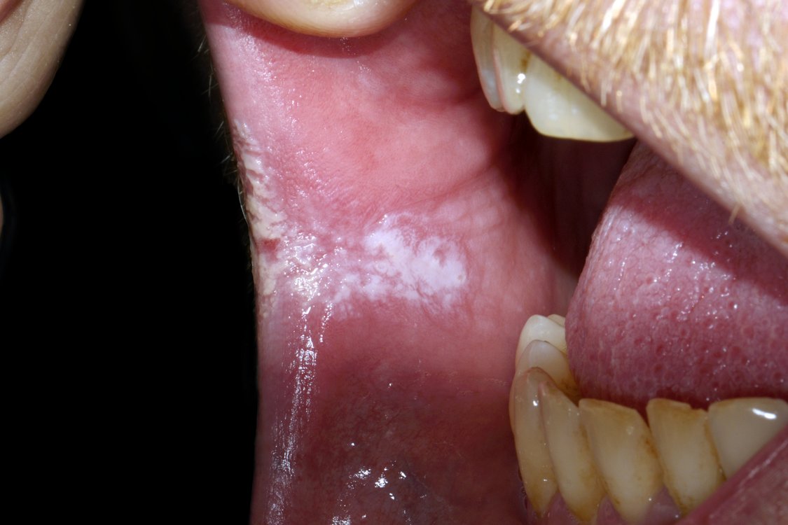
White patch on left buccal mucosa. Biopsy showed early squamous cell carcinoma. The lesion is suspicious because of the presence of nodules Nodular leukoplakia in right commissure. Biopsy showed severe dysplasia
The annual malignant transformation rate of leukoplakia rarely exceeds 1%, i.e. the vast majority of oral leukoplakia lesions will remain benign.A number of clinical and histopathologic features are associated with varying degrees of increased risk of malignant transformation, although other sources argue that there are no universally accepted and validated factors which can reliably predict malignant change. It is also unpredictable to an extent if an area of leukoplakia will disappear, shrink or remain stable.
• Presence and degree of dysplasia (mild, moderate or severe/carcinoma in situ). While the degree of dysplasia has been shown to be an important predictor of malignant change, many have challenged its use due to the low predictive value from the lack of objectivity of grading dysplasia. While 10% of leukoplakia lesions show dysplasia when biopsied, as many as 18% of oral lesions undergo malignant change in the absence of dysplasia.
• Leukoplakia located on the floor of the mouth, the posterior and lateral tongue, and the retromolar areas (the region behind the wisdom teeth) have higher risk, whereas white patches in areas such as the top surface of the tongue and the hard palate do not have significant risk. Although these “high risk” sites are recognized, statistically, leukoplakia is more common on the buccal mucosa, alveolar mucosa, and the lower labial mucosa. Leukoplakia of the floor of the mouth and tongue accounts for over 90% of leukoplakias showing dysplasia or carcinoma on biopsy. This is thought to be due to pooling of saliva in the lower part of the mouth, exposing these areas to more carcinogens held in
suspension.
• Red lesions (erythroplasia) and mixed red and white lesions (erythroleukoplakia/”speckled leukoplakia”) have a higher risk of malignant change than homogeneous leukoplakia.
• Verrucous or nodular areas have a higher risk.
• Although smoking increases risk of malignant transformation, smoking also causes many white patches with no dysplasia. This means that statistically, white patches in non-smokers have a higher risk.
• Older people with white patches are at higher risk.
• Larger white patches are more likely to undergo malignant transformation than smaller lesions.
• White patches which have been present for a long period of time have a higher risk.
• Persons with a positive family history of cancer in the mouth.
• Candida infection in the presence of dysplasia has a small increased risk.
• A change in the appearance of the white patch, apart from a change in the color, has a higher risk. Changes in the lesion such as becoming fixed to underlying tissues, ulceration, cervical lymphadenopathy (enlargement of lymph nodes in the neck), and bone destruction may herald the appearance of malignancy.
• White patches present in combination with other conditions that carry a higher risk (e.g. oral submucous fibrosis), are more likely to turn malignant.
• Although overall, oral cancer is more common in males, females with white patches are at higher risk than men
FAQ
Leukoplakia is a condition characterized by white patches or lesions that develop in the mouth's mucous membranes. While the exact cause is not always clear, leukoplakia is often associated with chronic irritation, such as tobacco use, alcohol consumption, or poorly fitting dentures. Regular dental examinations are crucial for early detection and management.
Diagnosis of leukoplakia typically involves a thorough examination of the mouth by a dental professional. In some cases, a biopsy may be necessary to confirm the diagnosis and rule out other potential concerns. Advanced imaging techniques may also be utilized for comprehensive evaluation and treatment planning.
What are the treatment options for leukoplakia? Treatment for leukoplakia depends on various factors, including the lesion's size, location, and underlying cause. In many cases, the primary approach involves removing the source of irritation, such as quitting smoking or addressing oral hygiene habits. Surgical intervention or laser therapy may be recommended for larger or persistent lesions. Regular follow-up appointments are essential to monitor progress and adjust treatment as needed.
While leukoplakia itself is not cancerous, it can sometimes indicate an increased risk of oral cancer, especially if the lesions are severe or persist despite treatment. Regular monitoring and early intervention are essential to detect any potential malignancies promptly. Maintaining good oral hygiene practices and avoiding known risk factors can help reduce the likelihood of complications.